|
|
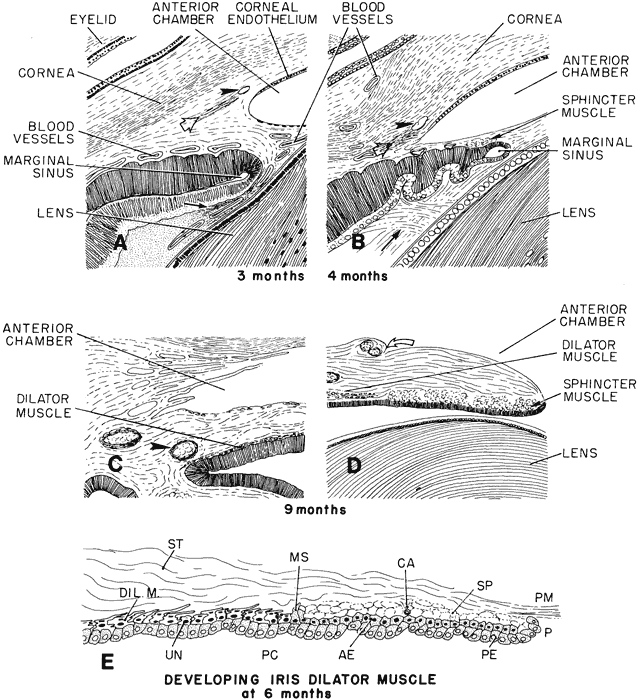
| Fig. 17. Schematic diagram of the progressive deepening of the angle; its relation to the neighboring tissues. A. At 3 months, corneal endothelium extends nearly to the angle recess: an incipient Schlemm's canal (arrowhead) and a more posterior scleral spur condensation (hollow arrow) appear to its left. Pigment epithelium of the forward growing ectodermal optic is indented by blood vessels. The secondary vitreous fibrils run parallel to its surface (arrow). This is the faisceau isthmique or marginal bundle of Druault. B. At 4 months, the angle recess has deepened and the endothelial lining has receded somewhat. There is a small aggregate of differentiating sphincter muscle fibers near the tip of the optic cup. Arrowhead points to Schlemm's canal. The condensed tissue just posterior to Schlemm's canal is the developing scleral spur (hollow arrow). Arrow points to the developing “tertiary vitreous” or zonular fibers. They originate from the nonpigmented ciliary epithelium and pass at right angles through Druault's bundle toward the lens capsule. C. The iris has grown and only its ciliary portion is presented. The angle recess has deepened and is occupied by loose connective tissue separated by many spaces. The dilator muscle of the iris has reached its root, which is still thick. Arrowhead points to the major arterial circle. D. Sphincter muscle is fully developed and is separated by connective tissue septa into several groups of cells. The collarette is represented as a surface stromal bulge containing two blood vessels (curved hollow arrow). E. Schematic diagram of the developing iris dilator muscle at 6 months. During the sixth month, dilator muscle fibers (Dil. M) start to differentiate from extensions of the anterior epithelial cells (AE) into the stroma (ST). These cells are located peripherally to the developing von Michel's spur (MS), which itself is a pigmented projection of the anterior epithelium, demarcating the posterior limit of the sphincter muscle (SP). In the developing dilator muscle, myofilaments within the elongating processes become arranged parallel to the stromal axis. Some undifferentiated anterior epithelial cells (UN) are present. In the sphincter, which had originated earlier from the same layer of anterior epithelial cells, connective tissue septa and a capillary (CA) start to grow between clumps of cells, but connective tissue has not yet invaded between the muscle cells and the anterior pigment epithelium beneath it. Eventually, the sphincter muscle bundles become completely separated from the anterior epithelium, whereas the dilator muscle sheet remains as the multilayered stromal projection of a part of this epithelium never separating from it. Therefore, the dilator muscle is not a separate cellular layer, but rather a partial myoid differentiation of cellular processes of the anterior neuroectodermal pigment epithelial cells. P, pupillary margin; PC, posterior chamber; PE, posterior epithelium; PM, pupillary membrane. |