|
|
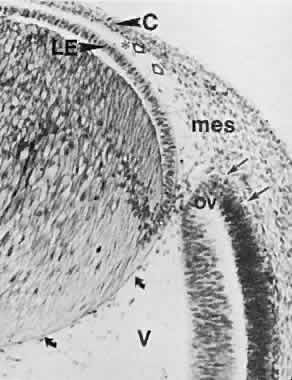
| Fig. 16. Embryo at 22 mm (approximately 7 weeks) showing relation of the anterior segment components (× 260). The two arrows indicate blood channels in the mesenchyme around the rim of the cup. Peripheral part of the pupillary membrane running from the mesenchyme in front of the optic cup (mes) to the anterior lens capsule outlines the incipient anterior chamber lying between it and the posterior surface of the cornea (hollow arrows). Asterisk is placed at the peripheral limit of the anterior chamber. Curved arrows point to capsula perilenticularis fibrosa. C, cornea; LE, lens epithelium; V, primary vitreous; ov, tip of the neuroectodermal optic cup. |