|
|
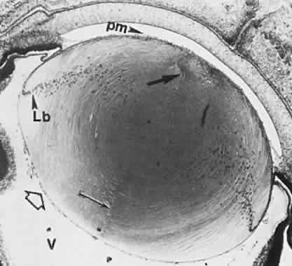
| Fig. 13. Lens at 65 mm (12-week fetus) in transverse section. Posterior suture (arrow) extends from the surface to the central, primary lens fibers (location of the embryonic nucleus). The triangular anterior suture (thick arrow) is indicated by an assembly of transversely cut fibers at the anterior pole. Posterior vascular lens capsule is indicated by hollow arrow. The nucleated area is the location of the secondary lens fibers. Lens bow (Lb) is formed by anteriorly migrating nuclei of newly formed lens fibers. pm, vessels of the pupillary membrane; V, vitreous (× 40). |