|
|
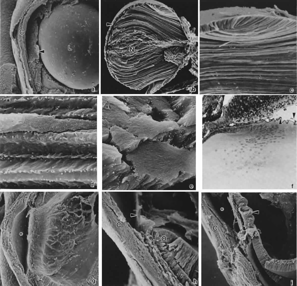
| Fig. 12. Formation of the lens and iridocorneal angle. A. Anterior segment at 8 weeks' gestation. The corneal stroma (C) and endothelium have formed. The dense pupillary membrane (arrow) fills much of the space within the anterior chamber. L, lens (× 100). B. Fractured lens at 7 weeks' gestation. Note embryonic nucleus (N) and anterior lens epithelium (arrow) (× 102). C. Higher magnification of (B) to illustrate secondary lens fibers and lens bow (× 376). D. Longitudinal view of lens fibers illustrating interdigitations (× 706). E. Cross-section of lens fibers illustrating tightly apposed hexagonal arrangement (× 1012). F. Light microscopic view of lens bow and close proximity of lens equator with anterior margin of optic cup. Note the hyaloid vasculature surrounding the lens (arrows) (× 220). G. At 8 weeks' gestation, following removal of the lens and the pupillary membrane, the anterior chamber can be visualized (× 103). H. Higher magnification of (G). The edge of the pupillary membrane can be seen (arrow) as well as the anterior margin of the optic cup (O) and the developing outflow pathways. The clefts visible in the limbal region canalize to form Schlemm's canal. C, cornea (× 220). I. At 13 weeks' gestation, there are immature ciliary processes located in the region of the future posterior iris (arrow). Differential growth with relative posterior movement of the inner optic cup, results in the ultimate mature conformations coinciding with exposure of the trabecular meshwork as described by Anderson (× 95). C, cornea; (B-E, courtesy of Dr. Kathy Sulik.) |