|
|
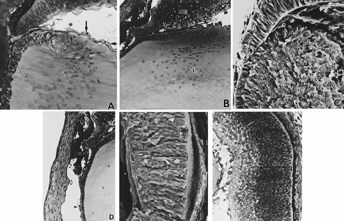
| Fig. 11. Formation of the lens fibers; early retinal differentiation. A. Elongation of the lens fibers located nearest to the neural retina forms the embryonal lens nucleus (L) and obliterates the lens vesicle cavity. The endothelial cells that form the tunica vasculosa lentis are indicated by arrows (× 392). B. Formation of the secondary lens fibers is apparent as elongation of the epithelial cells at the equatorial lens bow. C, cornea; NR, neural retina; L, lens (× 270). C. Electron micrograph evaluation of the developing lens (L). LE, anterior lens epithelium, E, surface ectoderm (× 298). D. Corneal endothelium (open arrow) and stroma (C) are completely formed but the anterior iridial stroma and iridocorneal angle (*) structures are still immature and covered by the endothelium. The outer, pigmented layer of the optic cup (O), which forms the pupillary sphincter and dilator muscles, is in apposition to the cornea in the area of the future aqueous outflow pathways (*). The arrowhead indicates the capillaries of the anterior tunica vasculosa lentis. L, lens (× 407). E and F. The retina has segregated into an inner neuroblastic layer (IN) containing the primitive ganglion cells the axons of which form the nerve fiber layer (arrow), and an outer neuroblastic layer (ON) containing the primordia of the photoreceptors, retinal interneurons, and glial cells (E, × 430; F, × 316). PE, retinal pigmented epithelium. |