|
|
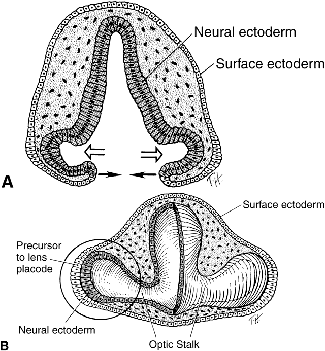
| Fig. 5. A. Drawing of a cross-section through forebrain and optic sulci of 24-day-old embryo. Note that the neural tube is still open. The optic sulci are lined by neural ectoderm (shaded cells), while the surface of the forebrain is covered with surface ectoderm (clear white cells). As the optic sulci (neural ectoderm) evaginate toward the surface ectoderm (hollow arrows), the edges of the brain vesicles move together to fuse, thus closing the neural tube (solid arrows). B. Drawing of a cross-section through a 26-day-old embryo at the level of the optic vesicle. Note that neural tube is closed, the surface ectoderm now lines the surface of the forebrain, and the neural ectoderm is completely internalized. The surface ectoderm cells overlying the optic vesicles enlarge to form the early lens placode. (Cook CS, Sulik KK, Wright KW: Embryology. In Wright KW [ed]: Pediatric Ophthalmology and Strabismus, pp 3–43. St Louis: Mosby, 1995.) |