|
|
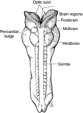
| Fig. 3. Drawing of 23-day-old embryo, dorsal view, showing partial fusion of the neural folds. Brain vesicles have divided into three regions: forebrain, midbrain, and hindbrain. Facing surfaces of the forebrain are lined with neural ectoderm (shaded cells), but the most of the embryo is now lined with surface ectoderm (clear white) because the neural groove has closed. On the inside of both forebrain vesicles is the site of the optic sulci. (Cook CS, Sulik KK, Wright KW: Embryology. In Wright KW [ed]: Pediatric Ophthalmology and Strabismus, pp 3–43. St Louis: Mosby, 1995.) |