|
|
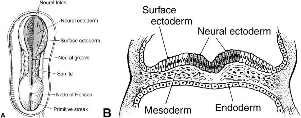
| Fig. 2. A. Drawing of dorsal view of a human embryo at 19 to 20 days' gestation. The neural plate transforms into two neural folds on each side of the neural groove. The neural groove in the middle of the embryo is shaded to represent neural ectoderm; the unshaded surface of the embryo is surface ectoderm. B. Cross-section of same embryo through the neural plate. Ectoderm in the area of the neural groove (shaded cells) has differentiated into neural ectoderm, whereas the ectoderm on each side of the neural groove is surface ectoderm (clear white cells) (Cook CS, Sulik KK, Wright KW: Embryology. In Wright KW (ed): Pediatric Ophthalmology and Strabismus pp 3–43. St Louis: Mosby, 1995.) |