|
|
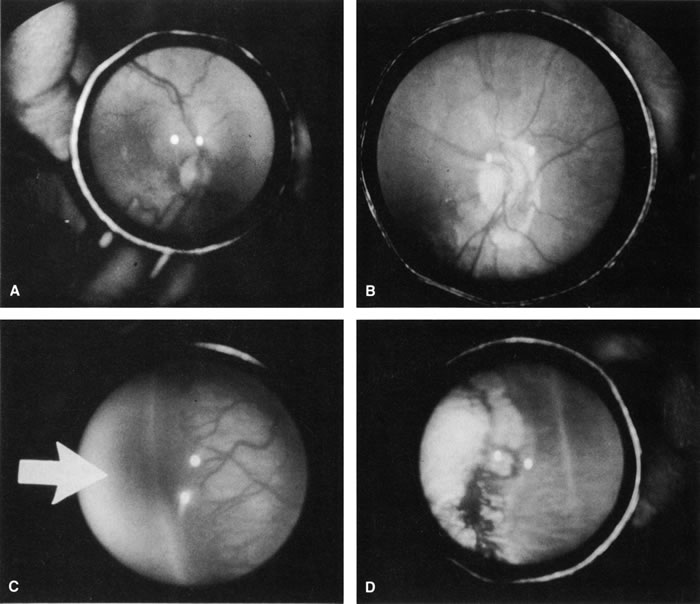
| Fig. 4. Preoperative (A) and postoperative (B) appearance of the posterior pole of an infant treated for threshold ROP with cryotherapy. Note the regression of plus disease after treatment. Preoperative (C) and postoperative (D) appearance of the retinal periphery of the same infant. Avascular zone (arrow). Note the regression of the extraretinal fibrovascular proliferation and the cryotherapy scarring. |