|
|
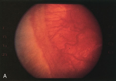
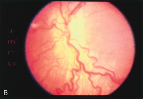
| Fig. 2. A. Clinical appearance of stage 3 ROP. This stage has growth of vessels with fibrous tissue out of the plane of the retina (extraretinal fibrovascular proliferation). B. Clinical appearance of plus disease. In the posterior pole, the retinal veins are engorged and tortuous. C. Two representative eyes that have reached threshold for treatment. The right eye (RE) has at least eight accumulative clock hours of stage 3 ROP. The left eye (LE) has at least five contiguous clock hours of stage 3 ROP. The thin line of ROP represents stage 1 or stage 2 disease, the broader sketched line signifies stage 3 disease. (From Cryotherapy for Retinopathy of Prematurity Cooperative Group: Multicenter trial of cryotherapy for retinopathy of prematurity. Preliminary results. Arch Ophthalmol 106:471–479, 1988. Copyright, Archives of Ophthalmology.) |