|
|
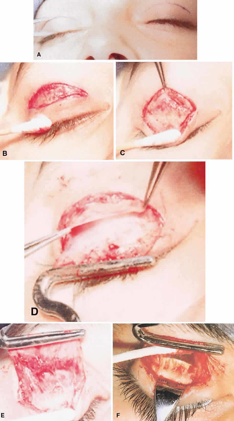
| Fig. 4. A. External levator resection, skin crease site measured and marked for lid crease incision. B. Skin and orbicularis incised, orbicularis layer undermined for short distance superiorly. C. Orbital septum and levator aponeurosis exposed as orbicularis layer is undermined. D. Orbital septum elevated from surface of levator aponeurosis, retroseptal fat contained. E. Levator aponeurosis grasped in ptosis clamp. Müller's muscle visible on undersurface of levator aponeurosis. Conjunctiva exposed above tarsus. F. Demonstration of subaponeurosis dissection with cotton-tipped applicator. The lid is held by an Ehrhardt's clamp that is visible through intact palpebral conjunctiva. |