|
|
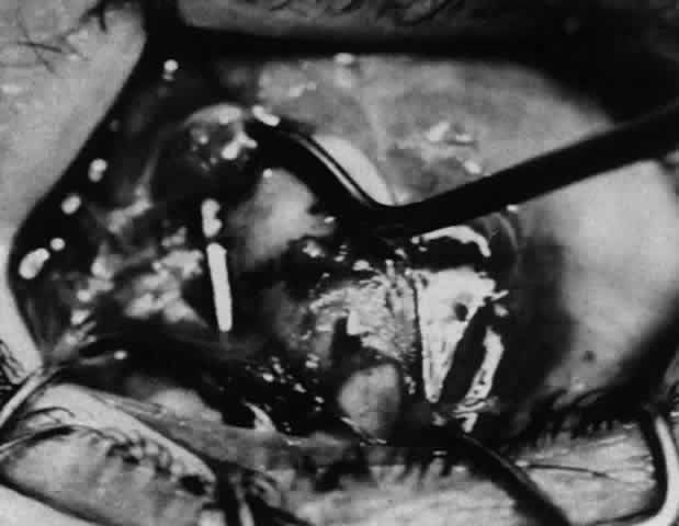
| Fig. 28. This intraoperative photograph demonstrates tissue that may simulate the insertion of the lateral rectus muscle. The cornea is to the right, just visible under the lid. The Jameson hook is under tissue that extends from the original insertion and the inferior oblique muscle. The true insertion is further posterior. The insertion of the muscle was found to be 17.5 mm posterior to the limbus. |