|
|
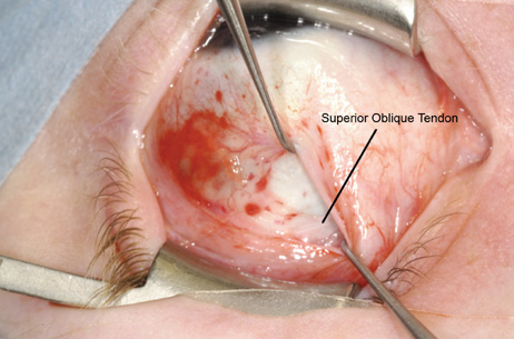
| Fig. 25. Although inferior traction is applied to the superior rectus with the large muscle hook, the lid speculum is removed and another Stevens hook is inserted into the wound to aid in exposure and visualization, exposing the parallel white fibers of the superior oblique tendon running beneath the superior rectus muscle, approximately 8 mm posterior to the superior rectus insertion. |