|
|
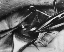
| Fig. 16. Two Stevens hooks are used to enlarge the opening in the check ligament, exposing the bare superior surface of the superior rectus muscle posteriorly. A Desmarres retractor is inserted through this opening to further attract Tenon's capsule and expose the nasal border of the superior rectus muscle, allowing direct visualization of the underlying parallel cordlike fibers of the superior oblique tendon (arrow). A small incision is made through the wispy capsule overlying the superior oblique tendon to bare the tendon fibers. |