|
|
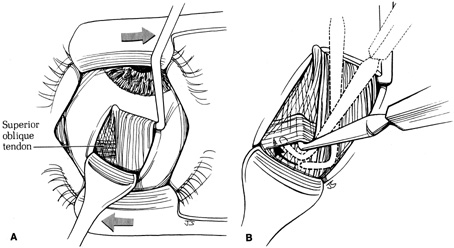
| Fig. 13. A. The Green muscle hook under the superior rectus muscle is pulled temporally and a Desmarres retractor moved nasally to improve visualization of the nasal border of the muscle. This maneuver exposes the nasal intermuscular septum, which covers and encases the parallel fibers of the superior oblique tendon as they pass beneath the superior rectus muscle. B. A Stevens muscle hook, with its tip pointing nasally, is moved posteriorly along the nasal border of the superior rectus muscle until the posterior margin of the cordlike superior oblique tendon is clearly identified. The tip of the hook is rotated posteriorly behind the tendon and passed toward the sclera anteriorly beneath the superior oblique tendon. (Del Monte MA, Archer SM: Atlas of Pediatric Ophthalmology and Strabismus Surgery. New York: Churchill Livingstone, 1993) |