|
|
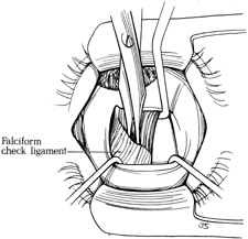
| Fig. 12. With the eye maximally depressed with the Green hook beneath the superior rectus muscle, the superior surface of the muscle is exposed with two Stevens hooks. The broad, sheet-like white falciform check ligament is exposed. This ligament fuses to the superior rectus muscle diagonally 8 to 12 mm posterior to its insertion. The ligament is opened centrally at its insertion with blunt Westcott scissors. (Del Monte MA, Archer SM: Atlas of Pediatric Ophthalmology and Strabismus Surgery. New York: Churchill Livingstone, 1993) |