|
|
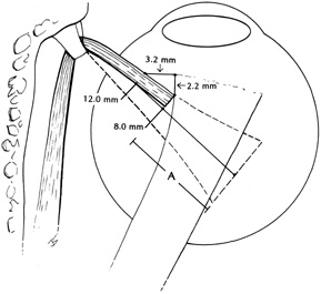
| Fig. 7. Landmarks for quantitating superior oblique recession as described by Romano and Roholt. The superior oblique tendon is secured with a double-armed 6-0 Vicryl suture and cut free from the sclera. This narrowed insertion is then recessed a measured amount (A) from the original insertion. The distance can be measured directly for small recessions (less than 6 mm). If it is recessed to a point 2.2 mm posterior to the medial border of the superior rectus recession, the net recession is 8 mm. If it is recessed to a point 3.2 mm nasal to the nasal border of the superior rectus muscle, it is recessed 12 mm, as shown. (Adapted from Romano P, Roholt P: Measured graduated recession of the superior oblique muscle. J Pediatr Ophthalmol Strabismus 20:136, 1983) |