|
|
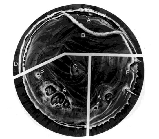
| Fig. 3. Top. Traumatic vitreous base traction. A. Upper nasal dialysis. B. Avulsed vitreous base. C. Dialysis at anterior vitreous base border. D. Tenting-up of retina and pars plana epithelium. Bottom left. Retinal breaks without vitreoretinal attachments. A. Large irregular breaks at the point of impact of blunt trauma. B. Small round holes in atrophic retina. C. Macular hole. Bottom right. Horseshoe and opercular tears. A. Of the equatorial retina. B. At the posterior vitreous base border. |