|
|
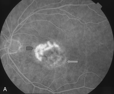
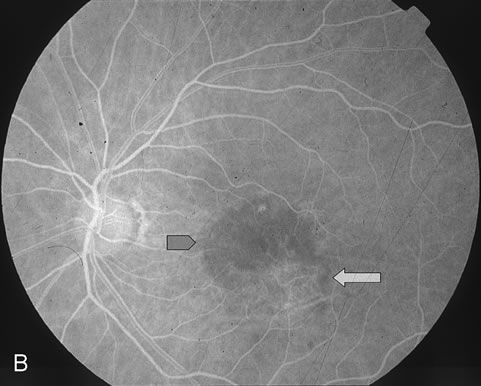
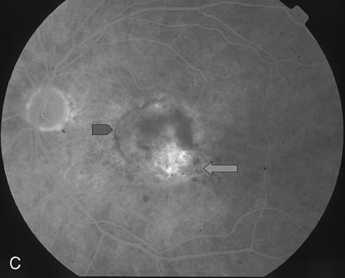
| Fig. 4. A. Mid-phase FA (1:23) demonstrates subfoveal hyperfluorescence corresponding to recurrent CNV (blue arrowhead) adjacent to an area previously treated with thermal photocoagulation (green arrow). B. Early-phase FA (28 seconds) 4 weeks status-post verteporfin-PDT demonstrates hypofluorescence (blue arrowhead) corresponding to the PDT treated area of recurrent CNV. C. Late-phase FA (3:40) 4 weeks status-post verteporfin-PDT demonstrates fluorescein staining in the area of prior thermal laser photocoagulation. There is a small rim of fluorescein staining around an area of hypofluorescence corresponding to the verteporfin-PDT treated area of recurrent CNV (blue arrowhead). |