|
|
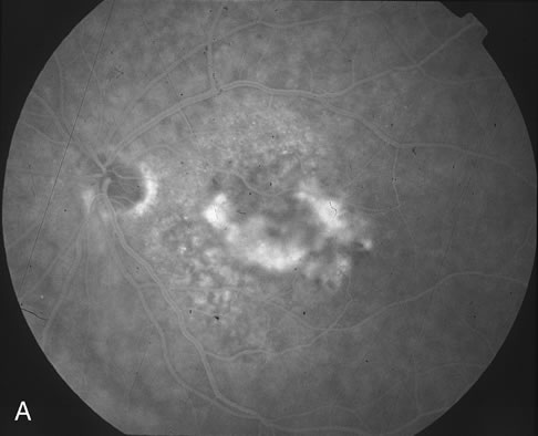
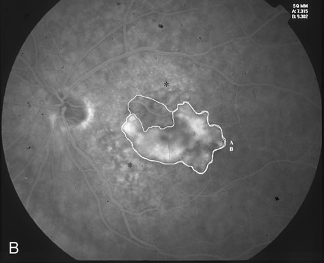
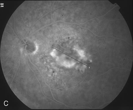
| Fig. 3. A. Fluorescein angiogram reveals predominantly classic CNV. B. Digital imaging outlines the classic component (A) and the entire lesion (classic and occult, marked as B). The area of classic component measures 7,315 square microns. The entire lesion measures 9,302 square microns. The areas marked with an asterisk correspond clinically to drusen. C. Greatest linear dimension (GLD) of the entire classic CNV measures 4,184 microns. |