|
|
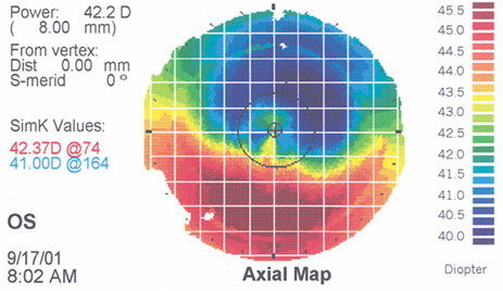
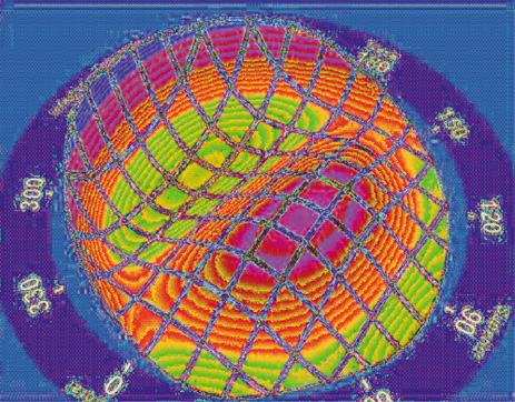
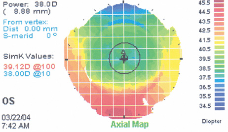
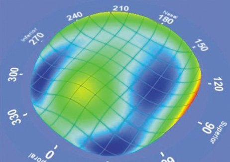
| Fig. 18. A. Corneal topography of a superiorly decentered myopic ablation. B. The three-dimensional wavefront shows a large amount of coma oriented vertically (90 degrees). C. After a wavefront-guided enhancement centered on the pupil, the corneal topography shows a marked improvement in centration. D. The postoperative wavefront is not perfectly flat but the coma is markedly reduced. |