|
|
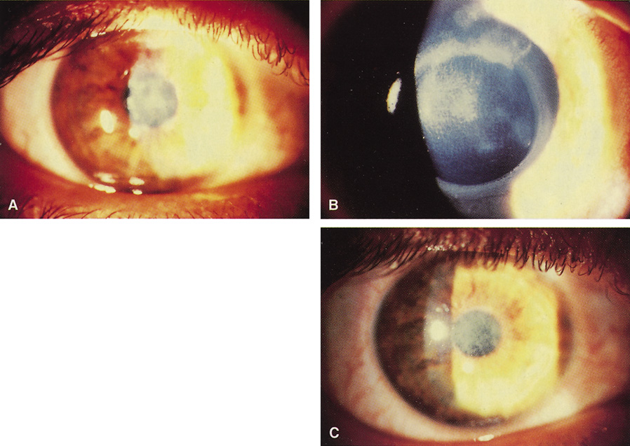
| Fig. 6. A. Herpetic anterior stromal scar before excimer phototherapeutic keratectomy. B. Appearance immediately after excimer phototherapeutic keratectomy, with reduction of the stromal scarring. The edge of the 4.5-mm–round ablation zone is evident. C. Three months after surgery, an oblique slit illumination shows some persistence of the deeper herpetic scarring plus the addition of reactive reticular haze. At this time, however, acuity was 20/30 versus 20/80 before surgery. |