|
|
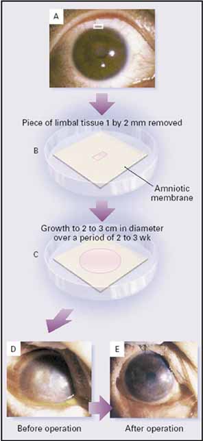
| Fig. 13 Schematic drawing of ex vivo expansion of limbal stem cells (SC). From the donor eye a small limbal biopsy is performed (A). One or two small explants (1 to 2 mm) are seeded on amniotic membrane in a culture medium (B). After 2 to 3 weeks, the explant yields epithelial outgrowth up to 2 to 3 cm of diameter (C). After the fibrovascular pannus is removed the explant is placed on the cornea (figures not shown). Preoperative picture shows limbal stem cell deficiency (LSCD) with central scar (D), and postoperative photo shows a smooth and clear cornea without corneal transplantation. (Reprinted from Tsai RJF, Li L-M, et al: Reconstruction of damaged corneas by transplantation of autologous limbal epithelial cells. N Eng J Med 343:86, 2000, with permission) |