|
|
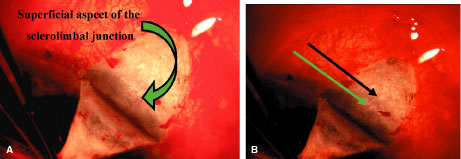
| Fig. 27. A. Deep sclerectomy with viscocanalostomy: completion of superficial scleral flap. The superficial flap should be uniform in thickness, leaving approximately 700 microns of subscleral tissue for the deep flap dissection. The flap requires dissection into clear cornea to ensure adequate exposure of Schlemm's canal and anterior tissues. The subscleral limbal landmarks start to become visible as the superficial flap is retracted. It is important to remember a standard trabeculectomy flap is much thicker and the limbal landmarks are in the deeper sclerolimbal junction. This is not the case in the Figure 26 in which the flap is only 300 microns. The green arrow indicates the anterior extent of the superficial sclerolimbal junction and does not contain the scleral spur, which is much deeper. B. Deep sclerectomy with viscocanalostomy: limbal structures identified with superficial flap. The anterior limit of the sclerolimbal junction is easily seen (green arrow) but the posterior limit is more difficult because the flap is very superficial. By slightly darkening the slide, the more posterior structures are more easily visible, the black arrow designates the deeper aspect of the sclerolimbal junction that should be the scleral spur. |