
|
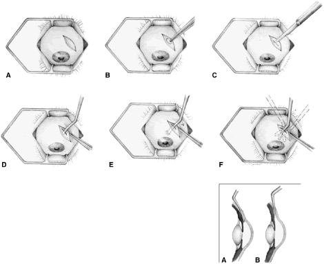
| Fig. 18. Cyclodialysis. A. An incision is made in the conjunctiva approximately 5 to 6 mm posterior to the limbus. B. Cautery is applied in the shape of an oval doughnut 5 mm long and 3 mm wide. C. An incision 4 mm long is made in the center of the oval. A traction suture may be placed. D. The tip of a cyclodialysis spatula is introduced into the space between the choroid and the sclera. E. As the tip is moved anteriorly, the heel of the spatula is depressed, and the tip is lifted up toward the microscope. A small bulge in the sclera results from the firm pressure employed. Inset. A. As soon as the tip is seen in the anterior chamber, it is retracted slightly and depressed posteriorly to avoid tearing Descemet's membrane. Inset. B. After the tip is in the anterior chamber, it is advanced anteriorly in a plane parallel to the iris. F. The spatula is swept to both sides. The pupil is observed carefully as the tip is moved posteriorly. The anterior ciliary vessels are meticulously avoided. Not shown is a keratostomy; this procedure is essential before the cyclodialysis can be performed. Keratostomy should be made 180-degrees opposite from the intended site of the cyclodialysis to permit irrigation of fluid into the cyclodialysis cleft. Air should be injected immediately after cyclodialysis to raise the intraocular pressure to approximately 30 mm Hg to keep the cleft well open and tamponade any bleeding. (Spaeth GL. Glaucoma surgery. In Spaeth GL (ed). Ophthalmic Surgery: Principles and Practice. Philadelphia: WB Saunders, 1990.) |