|
|
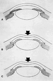
| Fig. 9. Indentation gonioscopy. A. The angle appears closed. However, the observer cannot determine whether this appearance is due to mere contacts between the iris and cornea or to actual adhesion. B. The goniolens has been pressed against the central cornea, displacing aqueous into the periphery and showing that the angle is open. C. Indentation gonioscopy displaces the iris posteriorly, showing peripheral anterior synechiae. (Schwartz LW. Diagnostic evaluation of the patient. In Spaeth GL (ed). Early Primary Open-Angle Glaucoma: Diagnosis and Management. Boston: Little, Brown Co, 1979.) |