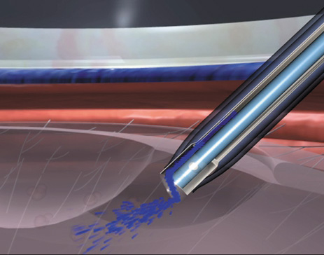
Fig. 19.
Diagram of the AquaLase tip showing fluid pulses disrupting the nucleus.