|
|
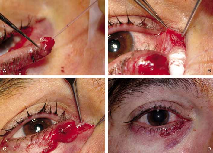
| Fig. 13 A–D. Monocanalicular stenting of canalicular laceration from Figure 12 in the office under local. A mini Monaka (FCI Ophthalmics, Marshill Hills, MA) is placed though the lateral portion of the lacerated canaliculus (A). The medial portion of the canaliculus is identified in the area of the canthal tendon (B) and the Silastic stent is fed into the lacrimal sac (C). The final appearance immediately on completion of canalicular and eyelid laceration repair (D). |