|
|
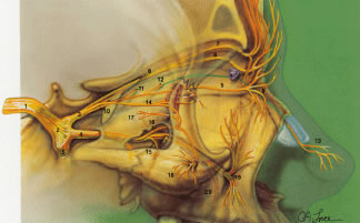
| Fig. 31. Sensory innervation to the orbit. Sensory nerves. (1) fifth cranial nerve; (2) trigeminal ganglion; (3) ophthalmic division of trigeminal nerve V1; (4) maxillary division of trigeminal nerve V2; (5) mandibular division of trigeminal nerve V3; (6) frontal nerve; (7) supraorbital nerve; (8) supratrochlear nerve (trochlea noted by purple); (9) infratrochlear nerve; (10) nasociliary nerve; (11) posterior ethmoidal nerve; (12) anterior ethmoidal nerve; (13) external or dorsal nasal nerve; (14) lacrimal nerve; (15) posterior superior alveolar nerve; (16) zygomatic nerve; (17) zygomatico-temporal nerve; (18) zygomaticofacial nerve; (19) infraorbital nerve; and (20) anterior superior alveolar nerve. (21) ciliary ganglion; (22) nerve to inferior oblique; (23) sensory root of ciliary ganglion; (24) long ciliary nerves; (25) short ciliary nerves. (From Zide BM, Jelkes G. Surgical Anatomy of the Orbit. New York: Raven Press, 1985.) |