|
|
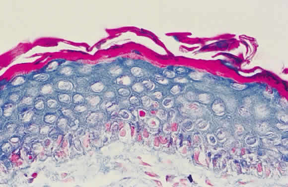
| Fig. 1. Histopathologic changes of conjunctival xerosis in a 25-year-old Indonesian woman. The epithelium is characterized by keratinization, a prominent granular cell layer, and distended squamous cells with large, open nuclei and prominent nucleoli. A mild mononuclear cell infiltrate, not apparent in this section, also was present. (H & E, × 700) (Sommer A, Sugana T, Djunaedi E, Green R: Vitamin A-responsive panocular xerophthalmia in a healthy adult. Arch Ophthalmol 96:1630, 1978) |