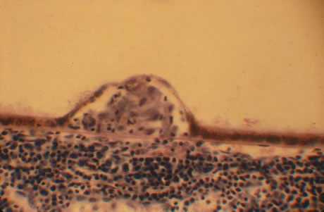
Fig. 59.
Histopathologic examination of a Dalen-Fuchs nodule shows inflammatory cells lying between Bruch's membrane and the attenuated retinal pigment epithelium.