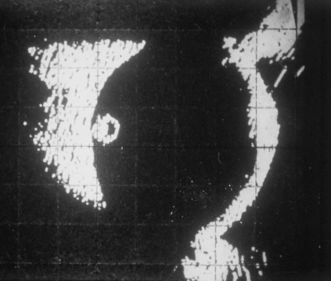
Fig. 41.
A B-scan ultrasonogram shows the cystic structure of the larva in the vitreous.