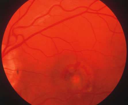
Fig. 30.
A fundus photograph of the posterior pole in case of presumed ocular histoplasmosis syndrome, showing peripapillary scarring and a choroidal neovascular membrane in the macula.