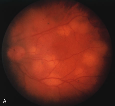
Fig. 10.
A
. The retinal involvement of
Pneumocystis carinii
is manifested by multifocal, yellow-white lesions.
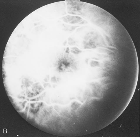
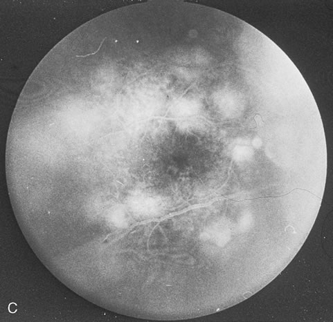
B
and
C.
On fluorescein angiography, the lesions fail to delineate in the early phase (B) but stain in the late phase (C).