|
|
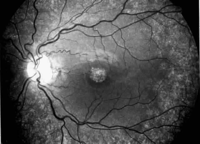
| Fig. 3. Cystinotic fundus changes in a 19-year-old patient, demonstrating a pale optic disc and numerous peripheral and macular small white spots at the level of the retinal pigment epithelium. Fluorescein angiography confirmed marked degenerative changes of the retinal pigment epithelium with macular, peripapillary, and peripheral window defects. |