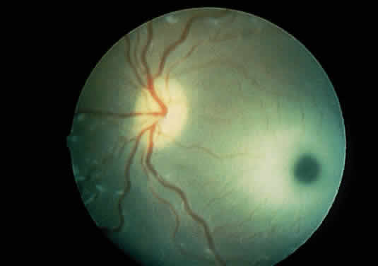
Fig. 21.
Posterior pole view of a patient with Tay-Sachs disease. The optic nerve head is atrophic, and there is opacification of the retina surrounding the foveal area, giving rise to a cherry-red spot.