|
|
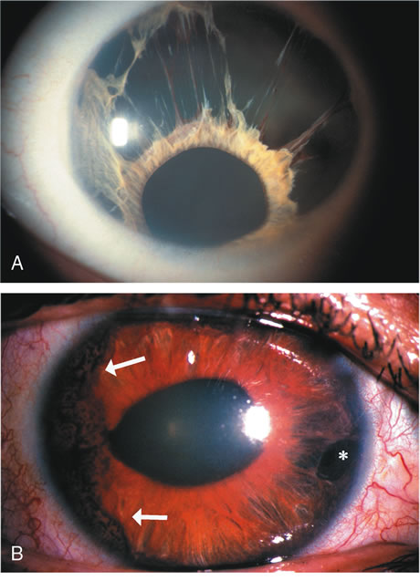
| Fig. 4. A. The iris in essential iris atrophy. Note the extensive stromal atrophy with strands of iris remaining superiorly. There is corectopia with preservation of the collarette and pupil. B. Cogan Reese syndrome. Note the melanocytic lesions (arrows) and the area of iris atrophy (*). (© University of Illinois at Chicago.) |