|
|
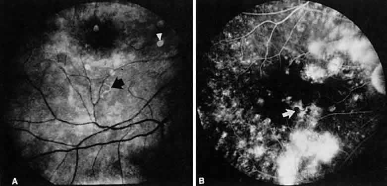
| Fig. 29. Diffuse unilateral subacute neuroretinitis. A. A coiled nematode (arrow) is seen in this red-free photograph just inferior to the right macula. Note the attenuation of the retinal arterioles. There are numerous subretinal grayish-white lesions involving the inferior portion of the right fundus. Five circular gray lesions are artifacts (arrowhead). B. Fluorescein angiography of the right posterior pole shows a hypofluorescent nematode (arrow) with multiple areas of hyperfluorescence due to diffuse retinal pigment epithelial changes. The nematode has now moved closer to the macula. |