|
|
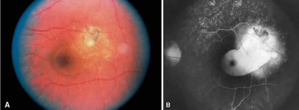
| Fig. 27. Congenital toxoplasmosis. A. Recurrent toxoplasmosis retinochoroiditis adjacent to an old scar with surrounding serous retinal detachment. B. Late venous phase angiogram showing discrete pooling of dye in the subretinal space due to an active toxoplasmosis lesion in the superotemporal macula. (Courtesy of Helmut Buettner, MD.) |