|
|
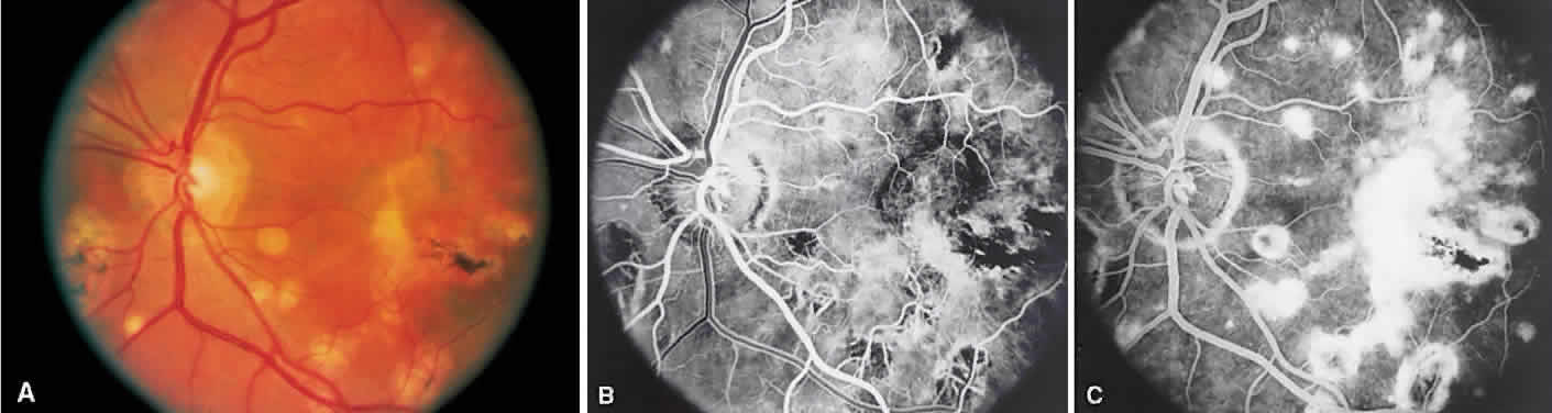
| Fig. 25. A Fundus photograph showing multiple punched-out chorioretinal lesions and disciform scar in the macula. B. Arteriovenous phase shows areas of hypofluorescence representing atrophic areas. C. Late venous phase photograph shows staining of the atrophic spots and leakage in the macula corresponding to the choroidal neovascularization. |