|
|
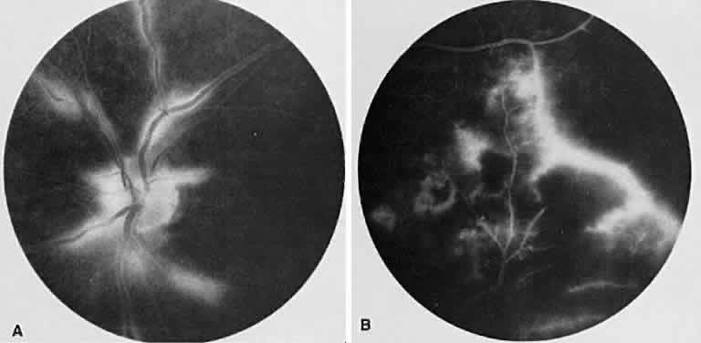
| Fig. 24. Eales' disease (retinal vasculitis). A. Angiography of the left disc shows staining of the retinal vessels, particularly the venules in the late venous stage. B. In the far periphery, there is staining of the peripheral vessels (particularly venules) just adjacent to an area of nonperfused retina. The hypofluorescent areas represent intraretinal hemorrhages. |