|
|
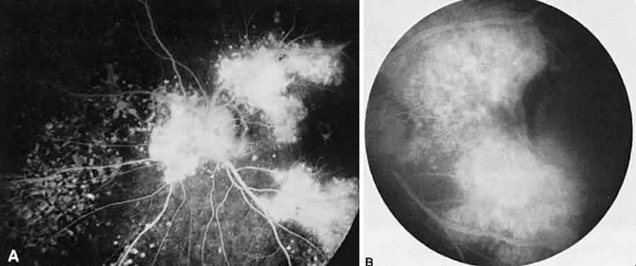
| Fig. 22. Syphilitic choroiditis. A. Fluorescein angiography of the left posterior pole shows peripapillary irregular mottling of the pigment epithelium with numerous areas of retinal pigment epithelial staining. B. Four months later, there is an increased subretinal hyperfluorescence temporal to the disc. The adjacent area of hypofluorescence represents subretinal blood. |