|
|
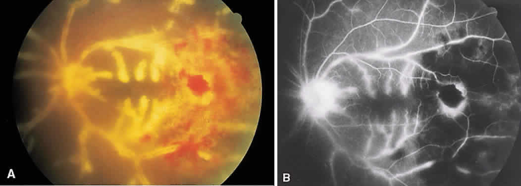
| Fig. 20. Frosted branch angiitis. A. Marked sheathing of retinal vessels with area of active retinitis and intraretinal hemorrhages is seen. B. Leakage of dye from the retinal venules representing retinal periphlebitis. Areas of hypofluorescence also are noted temporally corresponding to the retinitis. |