|
|
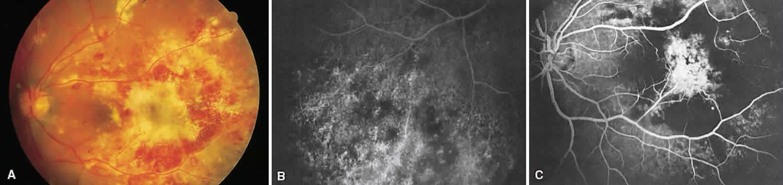
| Fig. 19. Cytomegalovirus retinitis. A. Fundus photograph of the left posterior pole shows necrotizing retinal changes with vascular sheathing and hemorrhages. B. Angiography shows large areas of hypofluorescence with a central region of multiple focal areas of staining within the lesion. Sheathed vessels stain but do not leak. C. Old cytomegalovirus retinitis. In an area of chronic retinitis, retinal pigment epithelium mottling is seen because of the necrosis. |