|
|
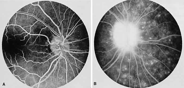
| Fig. 17. Multiple evanescent white-dot syndrome. A. Fluorescein angiography in the early venous phase shows a few punctate hyperfluorescent spots (retinal pigment epithelium window defects) around the disc. B. Late venous phase shows staining of more lesions and the optic disc. |