|
|
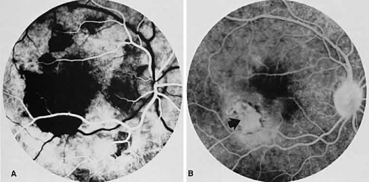
| Fig. 15. Subretinal neovascularization in serpiginous choroiditis. A. The arteriolar phase of the angiogram of the right eye shows a geographic center of choroidal hypofluorescence in the macula. There are additional areas of hypofluorescence superior to the right macular region. B. The venous phase of the angiogram shows a subretinal neovascular membrane just inferior and temporal to the macular zone (arrow). The hypofluorescence has decreased markedly in size compared with (A). The marked hypofluorescence masked the subretinal neovascular membrane initially. |