|
|
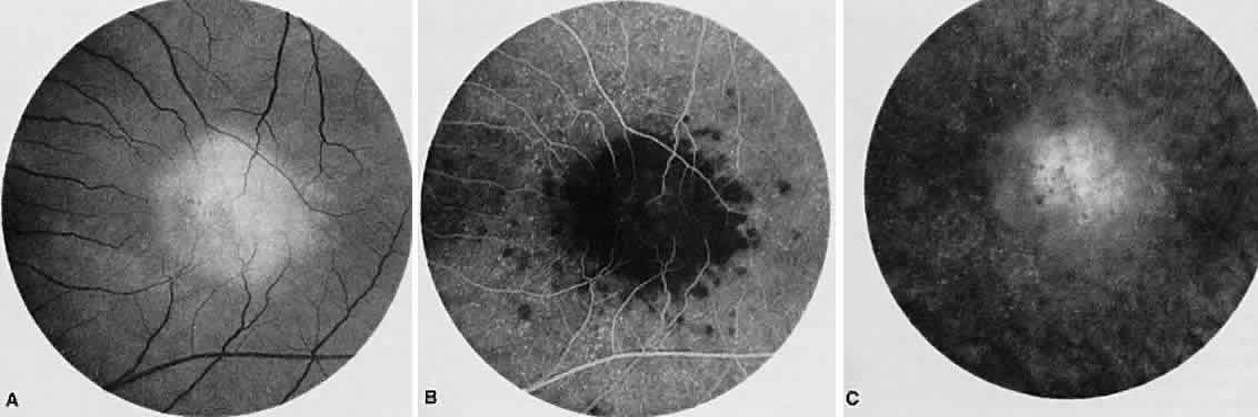
| Fig. 14. Macular serpiginous choroiditis. A. Red-free photograph of the left posterior pole shows the presence of a geographic lesion with diffuse borders at the level of the retinal pigment epithelium. B. Fluorescein angiogram of the left macular region shows a large area of choroidal hypofluorescence involving the macula. Other smaller hypofluorescent lesions surround the large lesion. Note pinpoint areas of hyperfluorescence representing drusen. C. The late venous phase shows diffuse staining of the left macular lesion. |