|
|
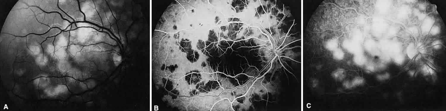
| Fig. 13. Acute multifocal posterior placoid pigment epitheliopathy. A. Red-free photograph of multifocal lesions at the level of the retinal pigment epithelium during the acute stage. B. Fluorescein angiography shows the multiple areas of hypofluorescence early in the arteriolar-venous phase. C. In the late venous phase, the hypofluorescent lesions have become hyperfluorescent. |