|
|
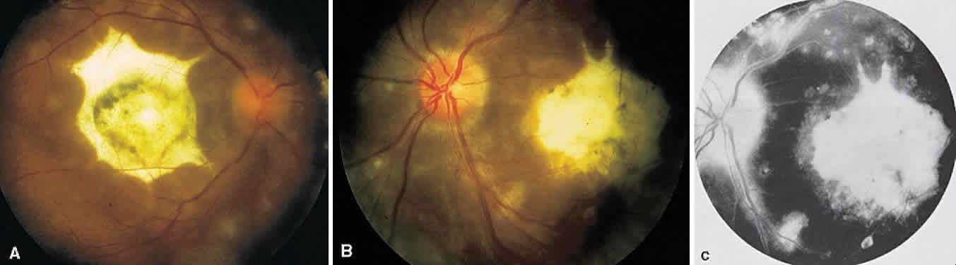
| Fig. 12. Multifocal choroiditis with subretinal fibrosis in a 26-year-old woman. A and B. Color photographs show hypopigmented lesions representing subretinal fibrosis involving both macular lesions. Multiple punched-out lesions surround the bands of fibrosis. C. Staining of the large stellate fibrous lesion can be seen in the left macula. There are multiple punched-out lesions above and below the macular zone. Leakage from the optic disc and its vessels can also be seen. |