|
|
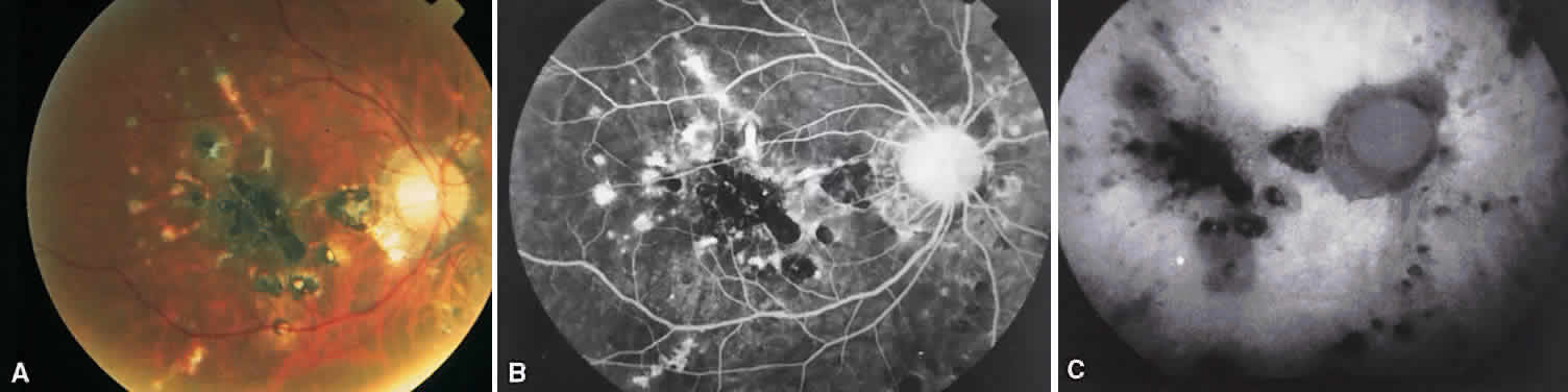
| Fig. 11. Multifocal choroiditis. A. Fundus photograph showing pigmentary disturbances. B. Multiple areas of hypofluorescence and hyperfluorescence representing chorioretinal scars with associated atrophic areas. C. Indocyanine green angiogram shows multiple areas of hypofluorescence around the disc, the macula, and the midperipheral fundus. Some of these areas are not visible clinically or on fluorescein angiogram. |